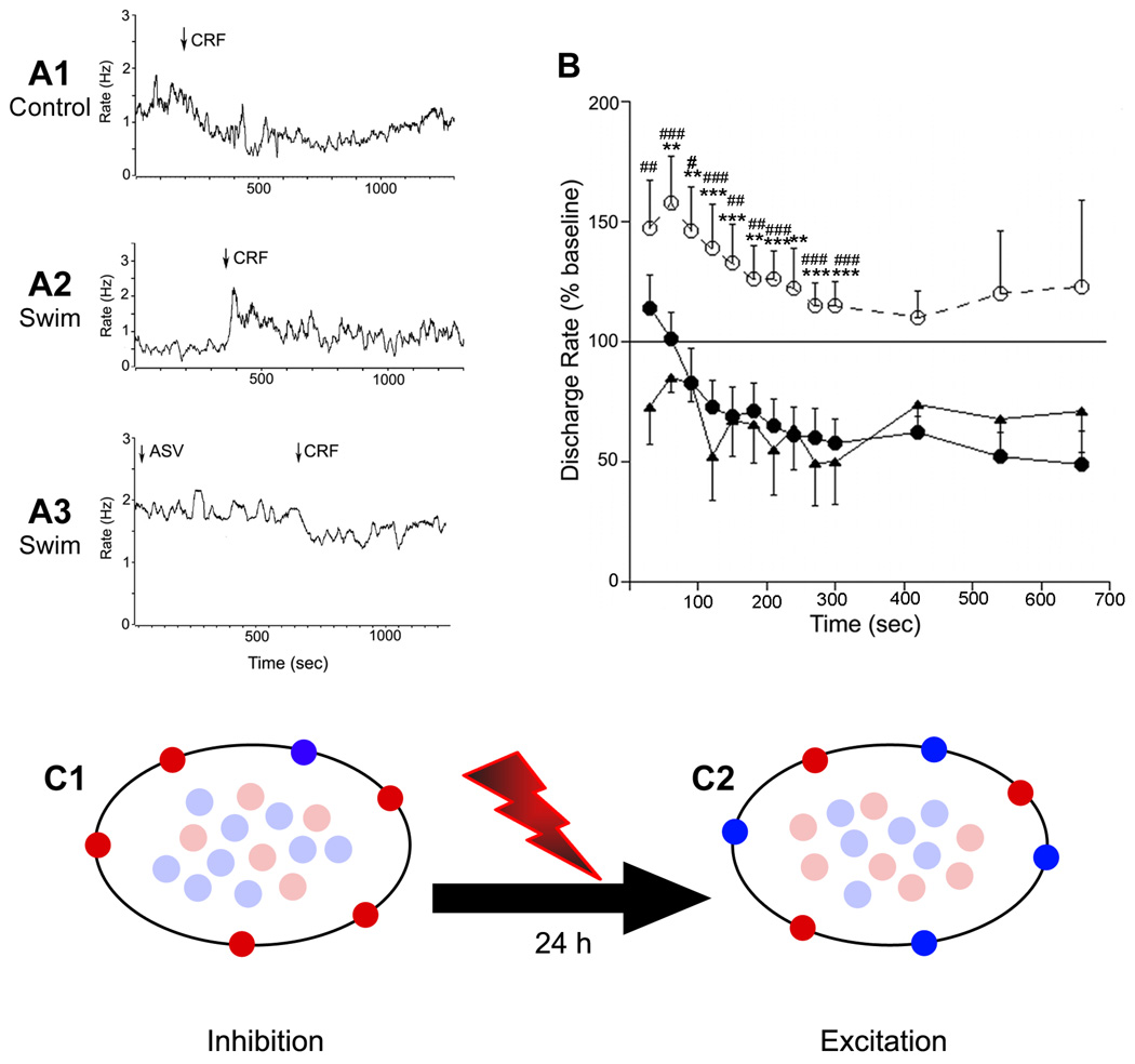
Figure 3. Swim stress results in a qualitative change in CRF effects on DR neuronal activity that favors CRF2 regulation.
A1–A3: Traces indicating the mean LC frequency of individual DR units recorded in an unstressed rat (A1), a rat exposed to swim stress 24 h prior to recording (A2) and a swim stress rat pretreated with antisauvagine-30 (A3). Arrows indicate the administration of CRF or antisauvagine (ASV); note the opposing effects produced by CRF in control vs. the swim stress rat. B: Time course of the effect of CRF on DR neuronal activity. The abscissa indicates the time after injection and the ordinate indicates the mean discharge rate expressed as a percentage of the mean rate determined over 200 seconds prior to injection. Shown are the mean effect of CRF in control rats (solid circles, n=11), swim stressed rats (open circles, dashed line, n=9) and swim stressed rats pretreated with antisauvagine-30 (solid triangles, n=6). Repeated measures ANOVA indicated a statistically significant effect of group (F(2,23)=8.4, p<0.002) and time (F(6,138)=7.0, p<0.0001 but no interaction (F(12,138)=1.0). Asterisks (*) indicate differences between control vs swim determined by Bonferroni post hoc test: **p<0.01; ***p<0.005; Number signs (#) indicate differences between swim vs ASV determined by Bonferroni post hoc test: #p<0.05; ##p<0.01; ### p<0.005. The mean basal discharge rates of cells in the three groups were 1.04±0.21 Hz, 1.94±1.02 and 1.18±0.05 for control, swim stress rats and swim stress rats that were administered ASV before CRF, respectively. These values were not different from one another (F(2,25)=1.2, p=0.3). C: Working model depicting how stress-induced redistribution of CRF receptors can result in a qualitatively different response to CRF. Schematic depicts cytoplasmic vs. plasma membrane localization of CRF1 (red) and CRF2 (blue) in unstressed rats (C1) and rats 24 h after swim stress (C2). In the unstressed condition CRF1 predominates over CRF2 on the plasma membrane and the neuronal response to CRF is inhibition. Twenty-four hours after swim stress the receptors are redistributed such that CRF2 predominates on the plasma membrane and the response to CRF switches to excitation.

