
Figure 2.
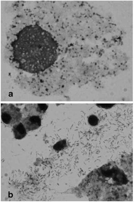
Appearance of Wolbachia in Giemsa-stained preparations. Samples were collected onto slides by centrifugation, dried, fixed in acetone, and stained with Giemsa stain as described in “Materials and Methods.” a Rounded forms of Wolbachia within a cell. b Extracellular, elongated forms. Photographs were taken with a ×100 objective.