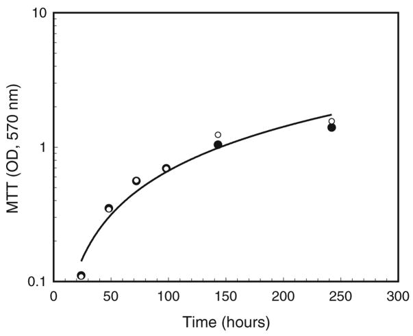
Figure 5.
Comparison of cell growth in the presence and absence of tetracycline. Cells were seeded at approximately 2×10⁵ per 35-mm plate in E-20 medium, allowed to grow at 28°C, 5% CO2, and assayed with MTT for 1 h at the indicated time points. Solid circles indicate Wolbachia-infected cells grown in the absence of tetracycline; the R2 value for the computer-generated trendline was 0.950. Open circles (note superimposition of some points) indicate cells grown in the presence of tetracycline from the time of plating. The R2 value for these data was 0.961.

