Figure 1.
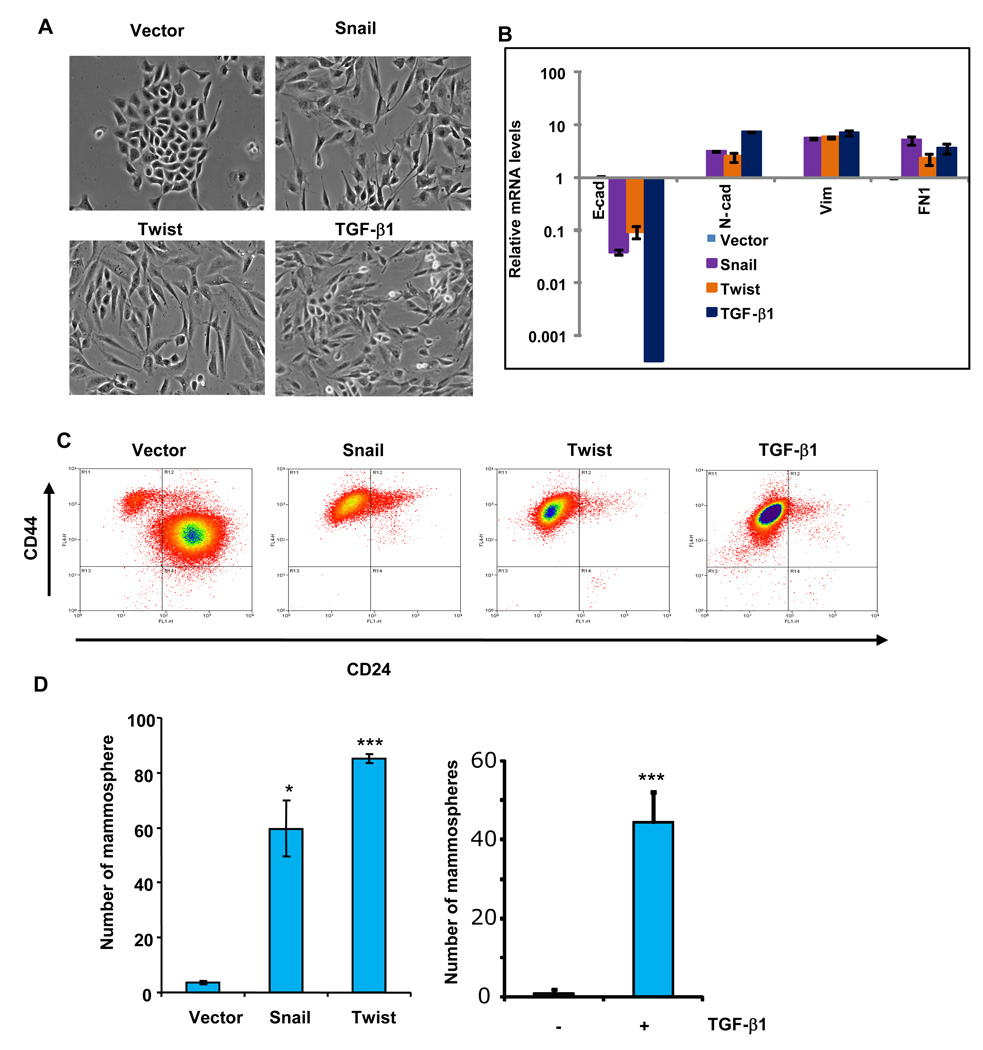
The epithelial-mesenchymal transition (EMT) generates cells with properties of stem cells. (A) Phase-contrast images of HMLE cells expressing Snail, Twist or the control vector, as well as HMLE cells treated with recombinant TGF-β1 (2.5 ng/ml) for 12 days (bottom right). (B) Relative expression of the mRNAs encoding E-cadherin, N-cadherin, vimentin, and fibronectin in HMLE cells induced to undergo EMT by the methods outlined in panel A, as determined by Real-time RT-PCR. GAPDH mRNA was used to normalize the variability in template loading. The data are reported as mean +/− SEM). (C) FACS analysis of cell-surface markers, CD44 and CD24, in the cells described in panel A. (D) In vitro quantification of mammospheres formed by cells described in panel A. The data are reported as the number of mammospheres formed/1,000 seeded cells +/− SEM, (* - P < 0.05; *** - P<0.001 comparing to the control).

