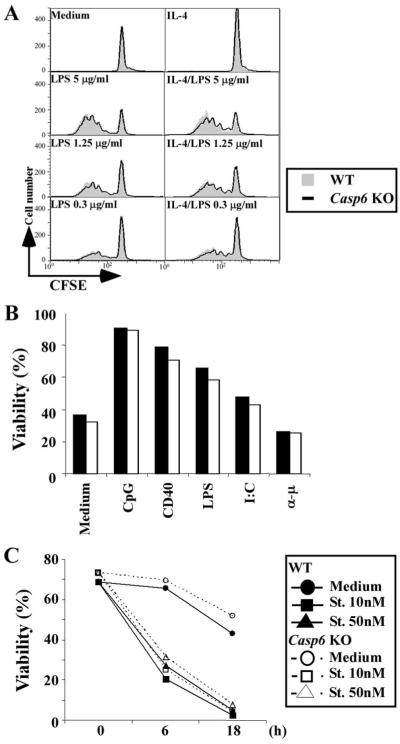
FIGURE 4.
B cell proliferation and cell death are not increased in Casp6 KO mice. A, Resting B cells were stained with CFSE, then stimulated with or without LPS (1 μg/ml) or LPS plus IL-4 (10 ng/ml) for 48 h. Cell division was measured on FL-1 using a FACScan. Gray areas show WT responses and thick lines show Casp6 KO responses. B and C, Purified resting B cells were treated with medium, CpG (1 μg/ml), CD40 (10 μg/ml), LPS (1 μg/ml), poly(I:C) (100 μg/ml), or anti-μ (10 μg/ml) for 48 h (B) or with staurosporine (St.; 10-50 nM) for 6 or 18 h (C). Cells were stained with MitoTracker ROS Red and analyzed with FACScan and CellQuest software. Percent viability is indicated for WT mice (closed bars or closed symbols) and Casp6 KO mice (open bars and open symbols). The data are representative of three independent experiments.

