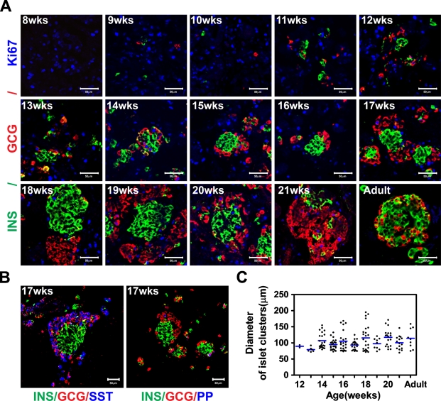
Figure 1.
Endocrine cells clustering during human pancreas development. (A) Immunostaining of human fetal and adult pancreas with anti-insulin (green), anti-glucagon (red), and anti-Ki67 (blue) antibodies. Numbers in the image indicate the gestational age in weeks (wks). (B) Seventeen-week fetal pancreas immunostained with anti-insulin (green), anti-glucagon (red), and anti-somatostatin (blue) or pancreatic polypeptide (PP) (blue). INS, insulin; GCG, glucagon; SST, somatostatin. (C) Diameter measurement of islet-like clusters. Clusters ≥ 70 μm were counted, and the size values were plotted. The lines indicate the mean ± SEM of the average diameter of the clusters. Bar = 50 μm.

