Figure 2.
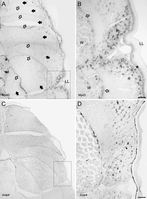
MyoD activity as revealed by IHC and bmp4 by ISH on cross sections (5 μm) of whole juvenile cod embedded in Technovit 9100 New. (A) MyoD activity is restricted to the dorsal and lateral extremes of the myotomes and a few cells deep inside the axial muscle along the myoseptum. (B) Magnification of stippled box in A visualizes MyoD activity in myosatellite cells of the white and red muscles. (C) Expression of bmp4 in white and red muscles is widespread and abundant in myocytes. (D) Magnification of stippled box reveals high bmp4 expression in both slow and fast fibers. No coloration was visible on the sections with a sense bmp4 probe control. And for IHC, no signals were present on control sections incubated with the secondary antibody only. The asterisk indicates pigmented melanocytes in the dermis and notochord and is not unspecific background. Closed arrowhead, myotome; open arrowhead, myoseptum; LL, lateral line; W, white muscle; R, red muscle. Bar = 40 μm.

