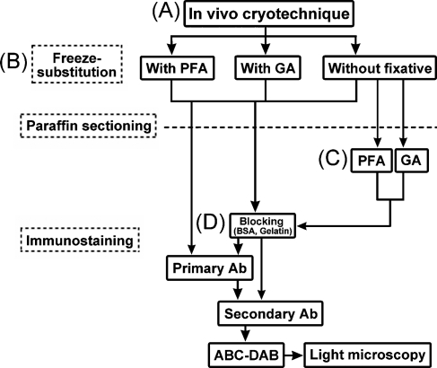
Figure 1.
A flow diagram of the preparation steps for the mouse eyeball tissues, as prepared by the in vivo cryotechnique (IVCT) (A) and freeze-substitution (FS) fixation for the glutamate (Glu) immunostaining. During the FS, paraformaldehyde, glutaraldehyde, or no fixative was added to acetone (B). Some thin sections of eyeball tissues without the chemical fixative during FS were treated with paraformaldehyde or glutaraldehyde (C). Before immunoreaction of the primary antibody, a common blocking treatment with bovine serum albumin (BSA) or fish gelatin was performed on the sections (D). PFA, paraformaldehyde; GA, glutaraldehyde; Ab, antibody; ABC-DAB, horseradish-avidin-biotin complex and diaminobenzidine reactions.

