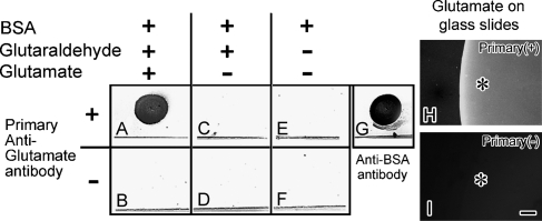
Figure 2.
Dot-blot analysis against variously prepared BSA with an anti-Glu antibody on nitrocellulose membranes (A–G). BSA-dotted membranes were treated with glutaraldehyde/Glu (A,B) or glutaraldehyde (C,D), or without treatment. BSA was assumed to be a binding dot pattern at the upper area of the line in each square. It was blocked with gelatin and incubated with (A,C,E) or without (B,D,F) the anti-Glu antibody. (G) Immunopositive control to show the binding of BSA as a dot pattern on the membrane with the anti-BSA antibody. Only after treatment with both glutaraldehyde and Glu (A), the immunoreactivity (IR) is obtained. (H,I) Glu-IR by attaching Glu to glass slides and immunostaining with (H) or without (I) the primary anti-Glu antibody (Primary) at higher magnification under a fluorescence microscope. Asterisks in (H) and (I) indicate the spot areas where the Glu was attached to glass slides. Bar = 50 μm.

