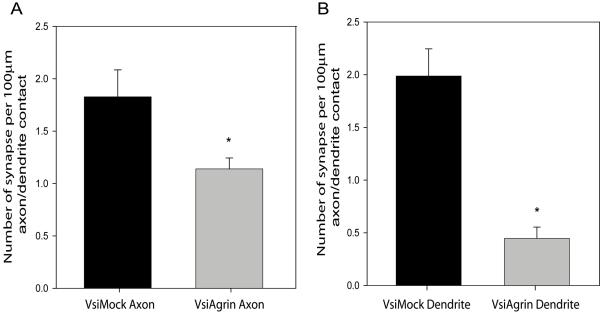
Figure 7. Depletion of Tm-agrin in dendrites reduces synapse density more than depletion of Tm-agrin in axons.
Synapse density was assayed in cultures such as shown in Figure 6, along segments of dendrites where it was clear that the only contact was between an infected axon and uninfected dendrite or vice versa. (A) Synaptic density along contacts with VsiAgrin-infected axons was reduced by 38± 2% relative to VsiMock-infected axons (p<0.01; n=16 segments from 8 neurons, 3 separate experiments), whereas (B) Synaptic density along VsiAgrin-infected dendrites was reduced by 79± 1% relative to VsiMock-infected dendrites (p<0.0001; n=14 segments from 8 neurons, 3 separate experiments). Synaptic density with VsiAgrin-infected axons was significantly different from synaptic density with VsiAgrin-infected dendrites (p<0.0001). Bars show mean ± SEM. Asterisks indicate values significantly different from VsiMock control.

