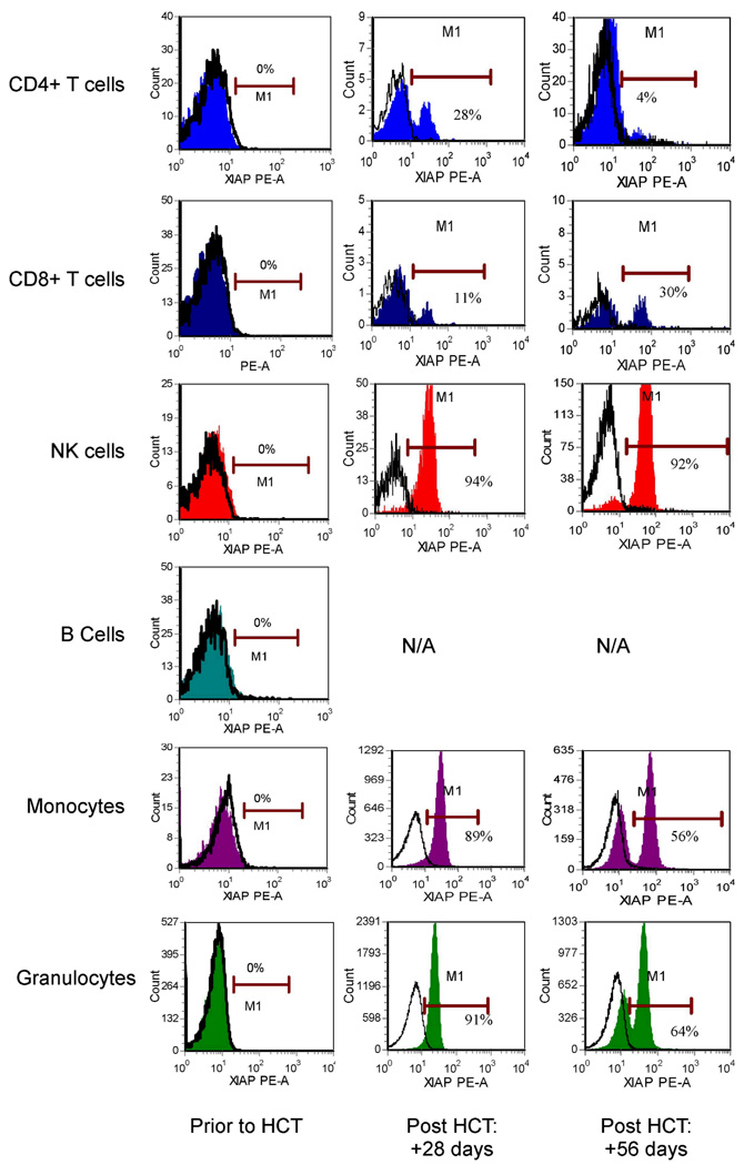
Figure 7.
Flow cytometric detection of XIAP in peripheral blood CD4+ T cells, CD8+ T cells, NK cells, B cells, monocytes and granulocytes from XIAP deficient patient 2, prior to and at +28 and +56 days following allogeneic hematopoietic cell transplant (HCT) using the clone 48 anti-XIAP antibody. Filled histograms represent XIAP staining, while open histograms represent control antibody. N/A=not applicable, as there was no significant B-cell reconstitution at these time points.

