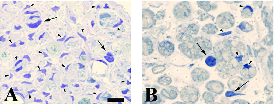
Figure 1.
Representative high-power views of 1µm plastic-embedded sections stained with toluidine blue. WT and transgenic TK animals received a crush lesion of the sciatic nerve and implantation of a minipump to deliver ganciclovir (GCV; 20 mg/kg/day) for the first seven days after surgery. Nerve segments distal to the crush lesion were harvested 7, 14, or 35 days post-injury. Panels are from the 14-day time point. A) WT animal. Note the numerous Schwann cell nuclei (identified by their characteristic dense staining and elongated or irregular morphology; arrowheads). A few white blood cells are also present (identified as larger moderately dark cells with rounded or oblong cell bodies and nuclei; arrows). B) Transgenic animal (TK). Compared to the WT animal note the paucity of Schwann cells but similar density of white blood cells. Size bar in A represents 10 microns for both panels. The quantification of such sections is shown in figure 2.

