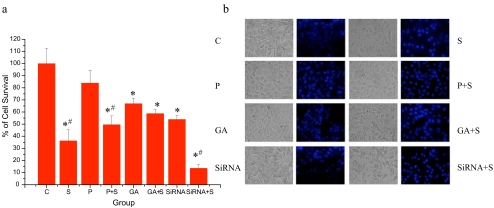
Fig. 1.
Oxidative preconditioning and GA treatment caused changes of cell viability and apoptosis in HepG2 cells during oxidative stress. a Effect of oxidative preconditioning and GA treatment on cell viability. Cell survival of HepG2 cells untreated (C) or treated with oxidative preconditioning (P: preconditioned with 2 h exposure to 50 μM H2O2, followed by 10 h recovery), geldanamycin (GA, 10 nM, 24 h), or small interference RNA of Hsp90 (siRNA) after oxidative stress (S: exposure to 500 μM H2O2 for 24 h), presented by MTT assay as percent of cell survival, where 100% survival is that of HepG2 cells that did not undergo oxidative stress (control). Results are from eight independent experiments (mean ± SE; *, #p < 0.05; asterisk each group vs control; number sign with oxidative stress vs without oxidative stress). b Effects of oxidative preconditioning and GA treatment on apoptotic morphology. Phase-contrast images (left and fluorescent images (right) were taken. The nuclear morphological changes were observed by Hoechst 33342 staining. In the control group, the nuclei of most HepG2 cells (>90%) were round and homogeneously stained. Cells treated with GA (10 nM) and oxidative preconditioning showed slightly increased cell numbers of typical morphologic features of apoptosis, such as chromatin condensation and fragmented fluorescent nuclei, which did not increased significantly compared to the directly oxidative stressed cells

