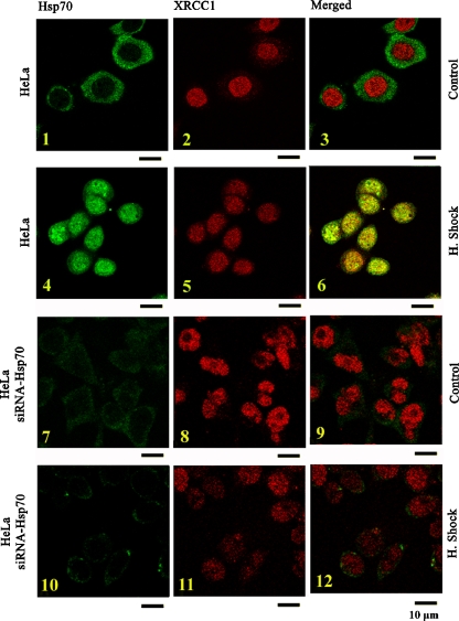
Fig. 4.
Subcellular distribution of endogenous Hsp70 and XRCC1 during control or heat shock. Confocal sections showing the simultaneous immunodetection of Hsp70 and XRCC1 in HeLa cells (insets 1–6) or in HeLa–siRNA–Hsp70 cells (insets 7–12). Cells were exposed, or not, to heat shock (90 min at 43.5 Subcellular distribution of endogenous Hsp70 and XRCC1 during control or heat shock. Confocal sections showing the simultaneous immunodetection of Hsp70 and XRCC1 in HeLa cells (insets 1–6) or in HeLa–siRNA–Hsp70 cells (insets 7–12). Cells were exposed, or not, to heat shock (90 min at 43.5°C and 90 min recovery at 37°C), fixed with 2% formaldehyde, and double immunofluorescence were applied using antibodies specific for Hsp70 and XRCC1. Bar, 10 μm

