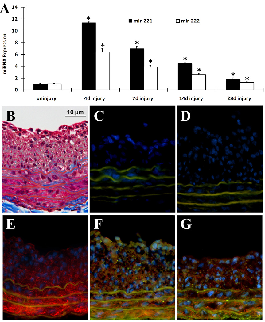
Fig. 1. Expression and distribution of miR-221 and miR-222 in balloon-injured rat carotid arteries.
(A). The time course changes of miR-221 and miR-222 expression determined by qRT-PCR. Note: n=6; *P<0.05 compared with uninjured control. (B). Representative Masson's trichrome staining. (C) Negative control (no SM α-actin antibody, no miRNA probe) for In situ hybridization and immunofluorescence. (D) Scrambled probe control 1 (no SM α-actin antibody, but had scrambled miRNA probe). (E) Scrambled probe control 2 (had SM α-actin antibody and scrambled miRNA probe). (F) In situ hybridization of miR-221 (dot green color), immunofluorescence of smooth muscle cell marker SM α–actin (red color) and cell nuclear staining by DAPI (blue color). (G) In situ hybridization of miR-222 (dot green color), immunofluorescence of smooth muscle cell marker SM α–actin (red color) and cell nuclear staining by DAPI (blue color). Note: Autofluorescence in the elastic laminae is demonstrated as green color, but is not dot green (C–G).

