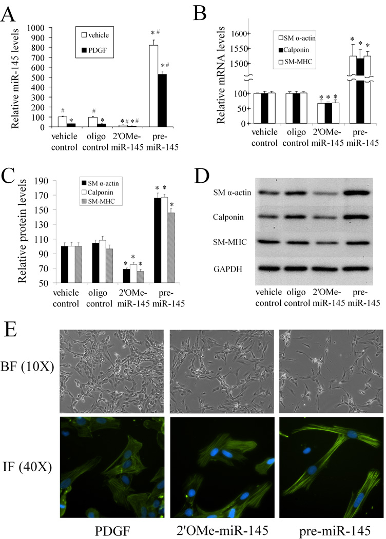
Figure 4. miR-145 modulates VSMC phenotype in vitro in cultured cells.
The VSMCs were pre-treated with vehicle, control oligo, 2'OMe-miR-145, or pre-miR-145 for 4h followed by PDGF or vehicle for 24 h. (A). Modulation of miR-145 expression by 2'OMe-miR-145 (100 nM) and pre-miR-145 (100 nM) in VSMCs with or without PDGF (20 ng/ml). n=6; *P<0.05 compared with oligo control group treated with vehicle. # p<0.05 compared with oligo control group treated with PDGF. (B). 2'OMe-miR-145 strengthened, whereas pre-miR-145 inhibited PDGF-mediated effects on VSMC maker genes as determined by qRT-PCR. n=6; *P<0.05 compared with oligo control. (C). 2'OMe-miR-145 strengthened, whereas pre-miR-145 inhibited PDGF-mediated effects on VSMC maker genes as determined by western blot. n=6; *P<0.05 compared with oligo control. (D). Representative western blots of VSMC differentiation marker genes. (E). Up panel: Representative morphological changes of primary cultured VSMCs treated with PDGF (20 ng/ml), 2'OMe-miR-145 (100 nM), or pre-miR-145 (100 nM) for 48 hours. Bottom panel: Representative immunofluorescence images of the VSMCs via anti-SM α-actin antibody (green color). Note: blue color is the cell nuclear staining by DAPI.

