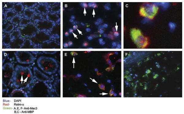
FIG 1.
Expression of Relm-α in DSS-induced colitis. Wild-type mice were treated with DSS for up to 8 days. Representative immunofluorescence microphotographs of Relm-α expression in the colons of wild-type control mice (A) and DSS-treated wild-type mice (B-F) are shown. Frozen sections were stained with anti–Relm-α (red; Fig 1, A-F), DAPI (blue; Fig 1, A-F), and either anti–Mac-3 (green; Fig 1, A, E, and F) or anti-MBP (green; Fig 1, B and C). A high-resolution image of double-stained MBP+/Relm-α+ cells is shown (Fig 1, C). Arrows indicate Relm-α+ cells (Fig 1, B-E).

