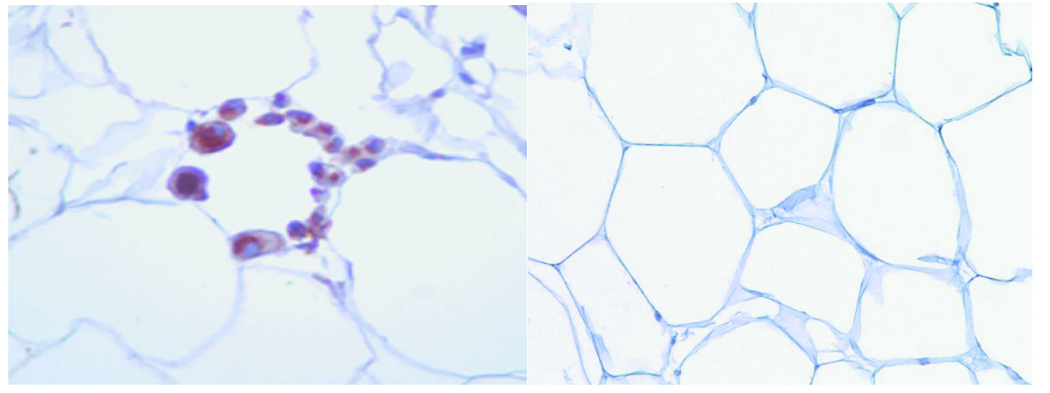
Figure 1.
Left panel: Representative light microscopic histology of CLS+ human subcutaneous abdominal fat. Aggregates of CD68 immunoreactive macrophages (brown color) are organized in crown-like structures around individual adipocytes as a hallmark of localized chronic inflammation in adipose tissue. Right panel: CLS− adipose tissue with absence of macrophage rings.

