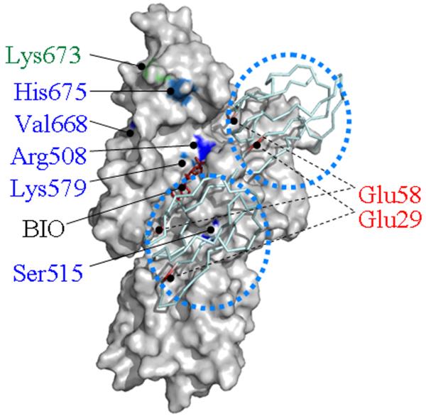
Figure 4.

Human HCS contains two potential docking sites for p67 near the biotin (BIO) and ATP binding sites. Two molecules of p67 are depicted as ribbon models and identified by using dotted circles. Functionally important amino acid residues in the central domain and C-terminal domain are highlighted in blue and green, respectively. Functionally important residues in p67 are depicted in red.
