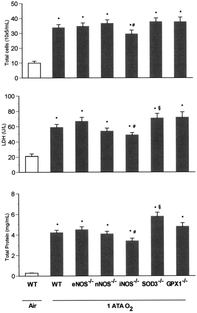
Fig. 1.
Pulmonary oxygen toxicity in 1 atmospheres absolute (ATA) O2. Wild-type (WT), eNOS−/−, nNOS−/−, iNOS−/−, SOD3−/−, and GPx1−/−mice (n = 11, 11, 10, 8, 9, and 8, respectively) exposed to normobaric hyperoxia (NBO2) for 72 h demonstrated severe increases in the measured indices of pulmonary injury compared with WT mice (n = 5) breathing room air (*P < 0.05 vs. WT in air). Scheffé’s post hoc test revealed that iNOS knockouts sustained significantly less pulmonary injury than did WT animals after exposure to NBO2 (#P ≤ 0.05 vs. WT in NBO2). By contrast, SOD3−/− mice showed more injury, with significant increases in bronchoalveolar lavage fluid (BALF) total protein and LDH activity (§P < 0.05 vs. WT in NBO2).

