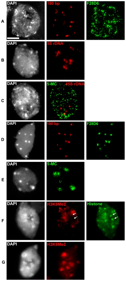
Figure 3. Cytogenetic characterization of Cvi-0.
FISH signals for the centromeric 180 bp [(A), red] and the subtelomeric 45S rDNA repeats [(C), red] are compact and located at chromocenters. Signals for the pericentromeric sequences 5S rDNA [(B), red] and transposon-rich BAC F28D6 [(A), green] are dispersed and outside heterochromatic regions. For comparison, in Col-0 both the centromeric 180 bp [(D), red] and BAC F28D6 [(D), green] are compact and located at the chromocenters. Immunolabeling of 5-Methylcytosine [(C), 5-MC, green] reveals a dispersed pattern in Cvi-0 compared to the clustered immunosignals in Col-0 [(E), 5-MC, green]. Note the absence of 5-MC signal on the Cvi-0 45S rDNA sequences [(C), red]. Immunolabeling of H3K9Me2 [(F), red] reveals the absence of this epigenetic mark on NOR chromocenters at the periphery of the nucleolus [(F), arrows] in Cvi-0, while all chromocenters are marked in Col-0 [(G), red]. Histone immunolabeling on Cvi-0 [(F), green] was carried out as control for histones. Each nucleus was counterstained with DAPI (first column). nu: nucleolus; Bar = 5 µm.

