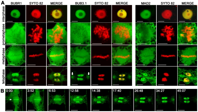
Figure 5. Subcellular localisation of BUBR1, BUB3.1 and MAD2 in tobacco cells undergoing normal mitosis.
(A) Single optical section of cells expressing BUBR1:GFP, BUB3.1:GFP and MAD2:GFP fusion constructs (green channel). Chromosomes in living cells were stained with SYTO 82 (red channel). In merged images, the yellow colour corresponds to the colocalisation of BUBR1:GFP, BUB3.1:GFP or MAD2:GFP with SYTO 82. By telophase, BUB3.1:GFP was detected in daughter nuclei (n) and in the midline at the cell periphery (arrow), forming a ring around the edge of the newly formed cell plate. (B) Selected frames from a fluorescence time-lapse analysis of the distribution of BUB3.1:GFP during cytokinesis. Single optical section of a cell expressing the BUB3.1:GFP fusion construct (green channel). After chromosome separation, BUB3.1 is localised along the midline of the anaphase spindle (arrowhead). During telophase, BUB3.1 is gradually transferred into the daughter nuclei. During phragmoplast extension from the centre to the periphery of the cell, BUB3.1 localises with the margin of the expanded phragmoplast. At the end of telophase, BUB3.1 is present at the cell periphery, forming a ring around the edge of the newly formed cell plate. This specific localisation at the phragmoplast midline disappeared when the newly formed cell plate completely separated the two daughter cells. At the end of cytokinesis, BUB3.1 was again concentrated in the nucleus. Time is in min:s. Bars, 10 µm.

