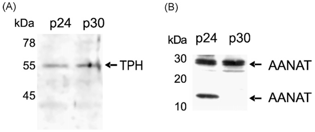
Fig. 2.
Detection of TPH (Panel A), AANAT (Panel B) immunoreactive proteins in human retinal pigment epithelium (RPE) cells. 50 µg of whole cell lysates of RPE passages 24 (p24) and 30 (p30), were resolved using SDS-PAGE. Immunodetection was performed using: sheep anti-TPH (1:300); rabbit anti-AANAT1–26 (1:5000) antibodies followed by an appropriate secondary antibody coupled to horseradish peroxidase (1:1000 or 1:5000, respectively). The presence of immunoprecipitates was visualized with Super Signal West Pico Chemiluminescent Substrate (Pierce). The chemiluminescent signal was acquired on a Fluor-S MultiImager and analyzed with Quantity One software (Bio-Rad).

