Figure 1.

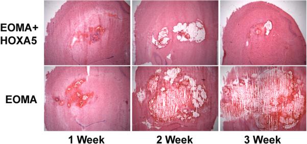
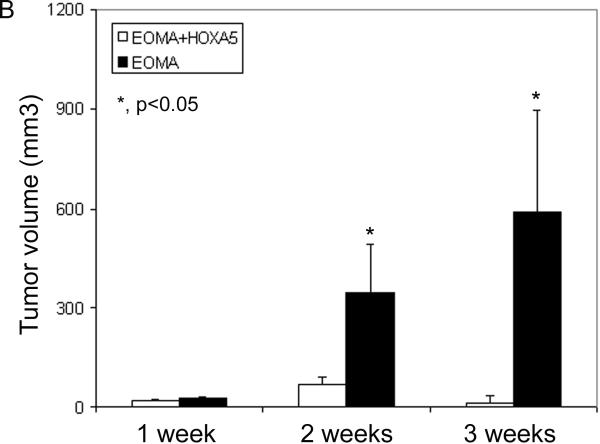
A) HoxA5 expression inhibits the growth of EOMA vascular lesions. HoxA5 (upper panels) or control transfected EOMA cells (lower panels) were stereotactically injected into the lateral caudo-putamen of adult CD1 mice and histologically examined 1, 2 and 3 weeks after injection. Photomicrographs show H&E staining of coronal sections at 1 (left side), 2 (middle) and 3 weeks (right side) following transplantation of EOMA+HoxA5 (upper panel) or EOMA cells (bottom panel) into the mouse brain. Tumors derived from HoxA5-expressing EOMA cells, were smaller than those induced by the control transfected EOMA cells. B) Bar graph shows the quantitation of the tumor volume in the EOMA+HoxA5 and EOMA groups at 1, 2, and 3 weeks after transplantation. Data are mean±SD, n=6 in each group. *p<0.05 for the difference between EOMA+HoxA5 vs. EOMA alone group for the same time point.

