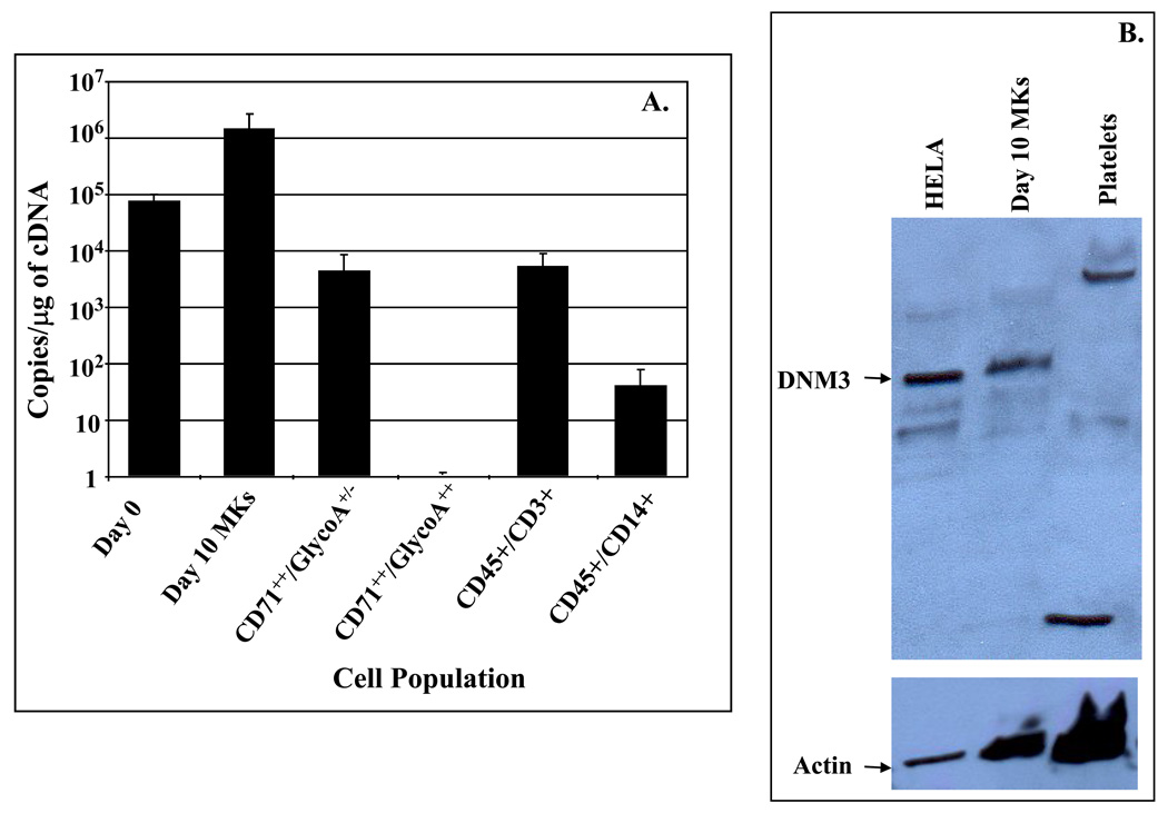
Figure 2. Quantitative RT-PCR (qRT-PCR) and Western Blot analysis of DNM3.
A) Cell populations tested by qRT-PCR include: Day 0 (i.e. uncultured CD34+/CD38lo BM cells); Day 10 MKs (i.e. culture derived MKs generated from CD34+/CD38lo after 10 days of culture with IL3, IL6, SCF & Tpo); CD71++/GlycoA+/− and CD71++/GlycoA+/− (i.e. isolated from volunteer donor marrow mononuclear fractions); and CD45+/CD3+, and CD45+/CD14+ cells (i.e. isolated from organ donor marrow);. Data are shown as mean±SD. B)Western Blot analysis was performed on Day 10 culture derived human MKs produced from umbilical cord blood CD34+ cells cultured for 10 days in media supplemented with IL3, IL6, SCF and Tpo. After 10 days of culture, the cells were harvested and stained with anti-CD41-PE. The stained cells were sorted by flow cytometry to obtain purified MKs, which were used to prepare protein lysates. Platelets were obtained from human peripheral blood. HeLa cell lysates were purchased and used as a positive control.

