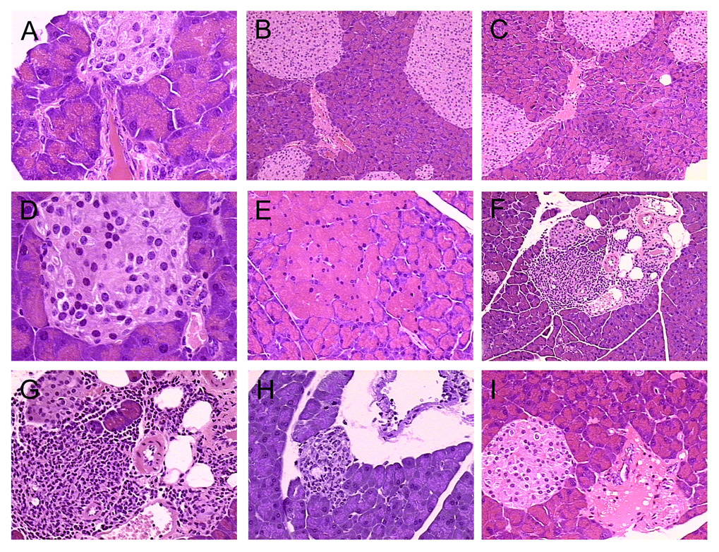
Figure 4.
Pdx-1-Aurora A transgenic mouse pancreatic lesions. (A) Most pancreas from these transgenic mice appeared normal with frequent mild dysplasia of ducts near islets, as evident in mouse 5. (B–D) Islet cell hyperplasia was evident in some of these transgenic mice including mouse 3 (B) and mouse 7 (C,D) . (E) Mild focal acinar hypertrophy was observed in mouse 6. (F) Mild fibrosis around an islet-ductal interface was detected in mouse 4. (G–H) Focal lymphocytic infiltration was observed around an islet-ductal interface in mouse 4 and a ductal lesion in mouse 9. (I) An odd fibrous lesion was evident near an islet-ductal interface in mouse 8.

