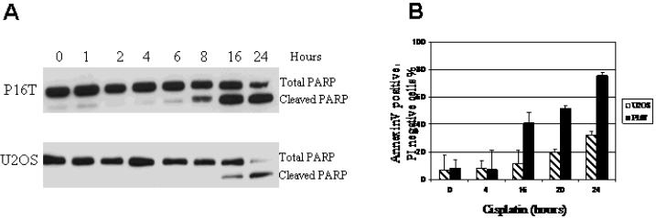
Figure 3. Cisplatin-induced apoptosis in OS cells.
P16T and U2OS cells were treated with 5μg/ml of cisplatin for the indicated time, and the cells with no drug added (0 hour) served as controls. The total and cleaved PARP was determined by Western blot analysis, annexin V/PI staining was assayed by flow cytometry. A: Increased cleavage of PARP is evident 8 hours following cisplatin treatment in P16T cells but not until 16 hours in U2OS cells. B: Significantly increased percentage of annexin V positive/PI negative cell population was obvious 16 hours following cisplatin treatment in P16T cells, but not until 20 hours in U2OS cells. In addition, the more robust increase in annexin V positive/PI negative cell population was observed in P16T compared to U2OS cells.

