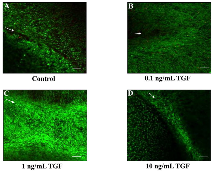
Fig. 5.
TGF-β1 increases cell accumulation at the interface of meniscal repair explants. Fluorescence confocal microscopy was utilized to visualize the cells in the meniscal repair interface. The green signal indicates live cells, while the red signal marks dead cells. Arrows mark the interface of the inner core and outer ring on all images. Representative images for each treatment are shown: control samples (A), 0.1 ng/mL TGF-β1 (B), 1 ng/mL TGF-β1 (C), and 10 ng/mL TGF-β1 (D). Scale bar = 100 μm.

