Figure 2.
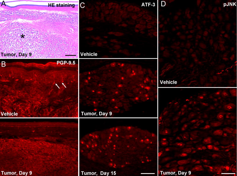
Nerve degeneration in the tumor bearing hindpaws. (A) Hematoxylin-eosin (HE) staining of hindpaw skin (plantar surface) 9 days after tumor inoculation. The tumor tissue (indicated with *) was located in the dermis. Scale bar, 400 μm. (B) PGP-9.5 immunostaining of hindpaw skin (plantar surface) reveals a loss of nerve fibers 9 days after tumor inoculation. Arrows indicated nerve fibers in the epidermis. (C) ATF-3 immunostaining indicates induction of ATF-3 in the nuclei of many DRG neurons after tumor inoculation. Scale bar, 100 μm. (D) pJNK immunostaining shows JNK activation in DRG neurons after tumor inoculation. Scale bar, 50 μm.

