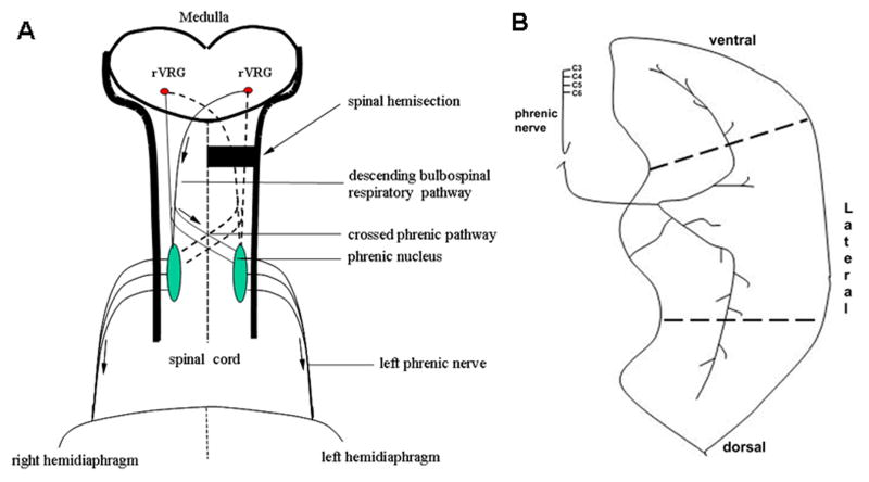
Figure 1.
A. Diagram of the crossed phrenic pathway in the adult rat. The pathway involves bilateral rVRG respiratory premotor axons with axon collaterals that cross the midline of the spinal cord. Arrows indicate the direction of respiratory impulses to the diaphragm after hemisection. B. Innervation of diaphragm muscle. The phrenic nerve primarily divides into rostral and caudal branches as it enters the diaphragm. EMG recordings were taken from 3 areas (ventral, lateral and dorsal) of the hemidiaphragm to assess the presence of the crossed phrenic activity immediately following C2 hemisection.

