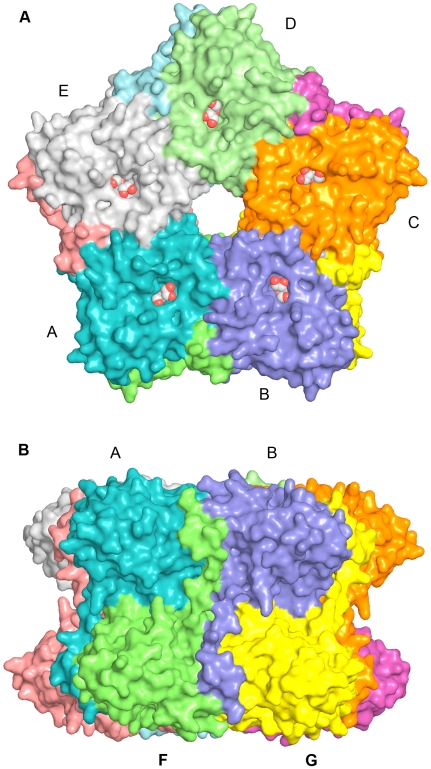
Figure 3. Structure of the LsrF decamer.
A. Surface representation of the LsrF decamer, viewed down the 5-fold symmetry axis, with each monomer a different color. The bound ligand (ribose-5-phosphate) is visible in the center of the (αβ)8-barrel, and is shown in ball-and-stick format. B. Perpendicular view of the LsrF decamer along a two-fold axis.

