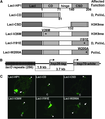
Figure 1.—
The LacI–HP1/lacO tethering system and localization of LacI–HP1 fusion proteins to polytene chromosomes. (A) The LacI DNA binding domain was fused to the amino terminus of HP1. The chromo domain (CD), hinge, and chromo shadow domain (CSD) of HP1 are indicated. The amino acid numbers are designated below each construct. Locations of amino acid substitutions are marked by asterisks and the function(s) affected by each mutation are given at right: D, dimerization; PxVxL, interactions with PxVxL partner proteins; and H3K9me, binding of dimethylated lysine 9 of histone H3. (B) The tagged hsp26 and hsp70-white transgenes are positioned 1.9 and 3.7 kb from the 256 lacO repeats, respectively. (C) Chromosomes were fixed, squashed, and stained with antibodies to LacI. LacI DNA binding sites at position 4D5 are denoted by an arrowhead, region 31 is indicated by an arrow, and the chromocenter is indicated by a “C.”

