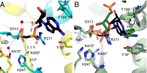
Fig. 2.
The AMPcP-binding site of human NAMPT. (A) Ribbon diagram showing the AMPcP binding mode with H247-unmodified human NAMPT. The 2 monomers are colored in yellow and cyan, respectively. AMPcP is shown in black and water molecules are depicted as red spheres with hydrogen bonds represented as olive dashed lines. The 5.3 Å green dashed line represents the distance from (Nδ1)H247 to the methylene α,β-bridge of AMPcP. (B) The structural overlap for PRPP·BzAM (substrates shown as green sticks with an overall representation of the structure in light green) and AMPcP complexes in nonphosphorylated human NAMPT (black stick representation of AMPcP with the corresponding structure in gray) is demonstrated. Produced with PyMol v 1.1.

