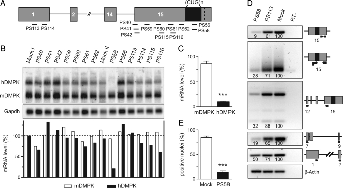
Fig. 2.
PS58-mediated silencing of expanded hDMPK transcripts in DM500 cells. (A) Location of target sites of AONs (black bars) along the hDMPK premRNA. (B) Northern blot analysis to detect ability of AONs to silence hDMPK mRNA in DM500 myotubes. Gapdh was used as loading control. Oligos were tested at least twice; a representative blot is shown. (C) PS58, directed at the (CUG)n segment, was the most successful AON (P < 0.001, n = 19). (D) PS58 activity was corroborated by semiquantitative RT-PCR analysis of individual segments of the hDMPK transcript (primers indicated with black arrows). Numerals in lanes indicate relative abundance to mock samples (set at 100), using β-actin for normalization. A representative result from 2 experiments is shown. (E) A reduction in the number of nuclei containing (CUG)n foci was observed after PS58 treatment of DM500 myoblasts (P < 0.001, n = 4).

