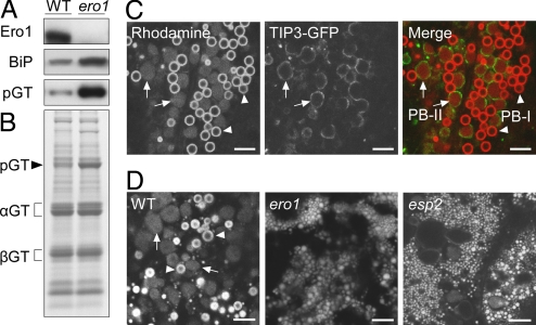
Fig. 5.
The RNAi knockdown of OsERO1 in seeds. Total extracts of the WT and ero1 seeds (17 DAF) were subjected to SDS/PAGE, followed by Western blot analysis with antibodies against OsEro1, BiP, and glutelin (A) or Coomassie Brilliant Blue staining (B); pGT, proglutelin; αGT, glutelin α subunit; βGT, glutelin β subunit. (C) Confocal fluorescence images of the subaleurone cells (17 DAF) expressing OsTIP3-GFP (PB-II membrane marker), stained with Rhodamine. (D) Confocal fluorescence images of Rhodamine-stained subaleurone cells (17 DAF) from the WT, ero1, and esp2 seeds. Arrowheads and arrows indicate PB-I and PB-II, respectively. (Scale bar, 5 μm.)

