
Figure 5.
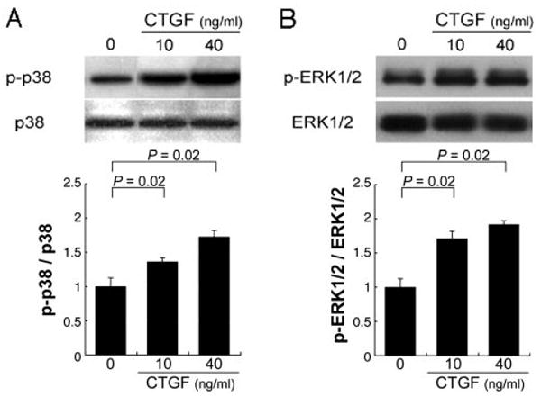
Activation of p38 (A) and ERK1/2 (B) MAP kinases by CTGF in ARPE-19 cells. Top: Western blot for phosphorylated and total levels of p38 and ERK1/2 in the ARPE-19 cells. Bottom: the graphs show the ratios of phosphorylated to total p38 (A) and ERK1/2 (B). p38 and ERK1/2 were significantly activated by CTGF. Statistically significant differences (P = 0.02) are seen, compared with controls.