
Fig. 1. Effect of R5020 and P on epithelial cell proliferation and organoid morphology.
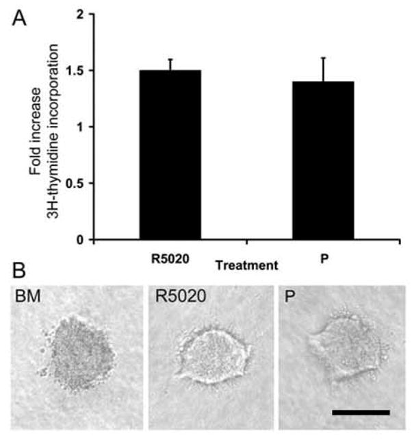
A. Mammary epithelial cells suspended in collagen I gels were cultured in BM, R5020 (20 nM), or P (20 nM). [3H] Thymidine incorporation into DNA was assayed after 3 d of culture. Each bar represents the mean ± SEM of triplicate values from a representative experiment. The data are expressed as the fold increase over the BM control. B. Organoid morphology was visualized in situ in collagen gels with the aid of an inverted microscope. Note the solid appearance of organoids from BM, whereas lumens are visible in R5020- and P-treated organoids. Scale bar = 100 μm.