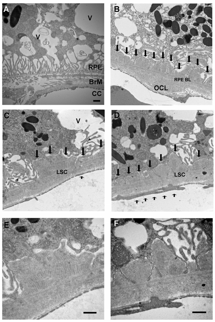
Figure 3.
Transverse Electron Microscopy of 12 month ApoB100 Mice Fed a Normal Diet. A. Marked ultrastructural derangement of the RPE with multiple cytoplasmic vacuoles (V), some of which contain membranous debris. The basal infoldings are fewer in number. An intercapillary bridge is seen in Bruch membrane separating two choriocapillaris lumens. B. Small basal laminar deposits replace lost basal infoldings (arrows) and an outer collagenous layer deposit (OCL) is identified. C–D. Truncated basal infoldings (*) are adjacent to a basal laminar deposit which contains long spacing collagen (LSC). Short arrow shows focal loss of choriocapillaris endothelial cell fenestrations. E–F. Higher magnification of C–D, respectively, showing long spacing collagen. RPE, retinal pigmented epithelium; RPE BL, RPE basal lamina; BrM, Bruch membrane; CC, choriocapillaris. Bar = 500 nm.

