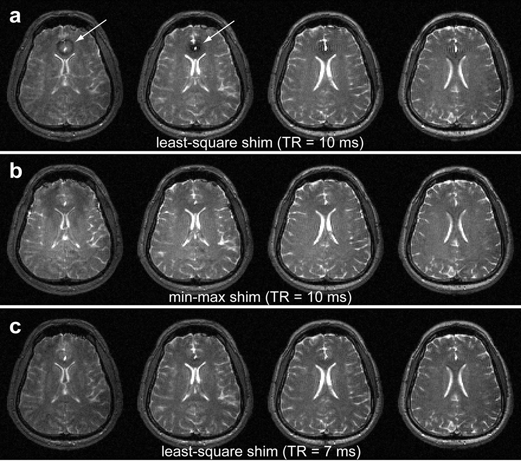
Figure 3.
Min-max shim result vs least-squares shim result. When the TR was 10 ms, the bSSFP images from the least-squares shim show the banding artifact (arrows) near the sinus area (a). The artifact disappeared when the shim was reconfigured to the min-max shim for the same TR (b). In the least-squares shim, the TR needed to be shortened to 7 ms to yield artifact free images (c).

