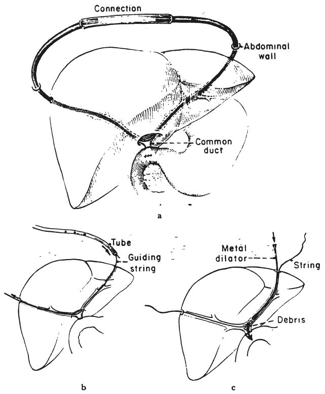
Fig. 1.
Technique of U tube placement. a, The U tube is in place with the ends joined by a connecting tube. b, To change the U tube, the Silastic tube is withdrawn with a trailing heavy silk suture, which is used to pull through another tube. c, The right and left ducts as well as the anastomosis can be obturated and dilated with a Bakes dilator.

