
Fig. 4. Vacuolar morphology of the tetR-VPS1 strain.
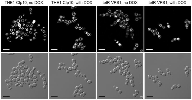
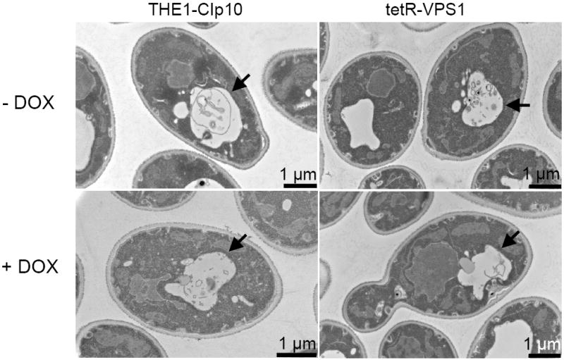
Vacuolar morphology was visualized by FM4-64 staining (A). A 10 μm bar is shown in either white or black. Thin-section electron microscopy images are shown in (B). A solid arrowhead is used to designate the vacuole. In either set of images, no major differences in vacuolar morphology were seen between the URA3-complemented wild-type strain THE1-CIp10 and the tetR-VPS1 strain, with or without doxycycline.