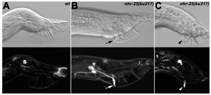
Fig. 3.
Loss of nhr-25 function leads to ectopic expression of mab-5 in ray 1. Nomarski images (top row) show male tail structures with sensory rays; below are confocal images of the mab-5::gfp activity. (A) In a wild-type adult male, none of the nine sensory rays show mab-5 expression; only autofluorescence of the spicule (s) and at the tail tip is apparent. (B,C) nhr-25(ku217) males display defective tail morphology (missing or fused rays) and abnormal mab-5::gfp expression that persists in ray 1 until the adult stage (arrowhead and arrow). Moreover, the ectopic mab-5 signal is also visible in cell bodies of the R1A, R1B and R1st (asterisk in B). Ray 1, which shows the mab-5 activity in C, is fused to ray 2 (arrowhead).

