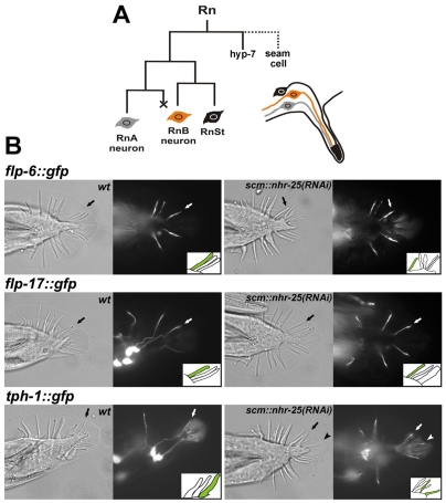
Fig. 5.
Ectopic rays in scm::nhr-25(RNAi) males exhibit ray-9 identity. (A) Lineage diagram of a ray cell group with two neurons and a structural cell. X represents programmed cell death. (B) Nomarski and fluorescence images illustrating the expression of three markers of specific RnB neurons. Schematic drawings (insets) of fluorescence images show expression of flp-6, flp-17 and tph-1 in T-seam-cell-derived rays. Arrows indicate wild-type signals in B neurons of the T-seam-cell-derived rays (R7B or R9B). flp-6::gfp is expressed in R2B (out of focal plane), R5B, R6B and R7B in the wild type (left). scm::nhr-25(RNAi) males (right) display flp-6::gfp signal in the same RnB neurons as wild-type males (R2B is out of the focal plane), but not in any of the ectopic rays. Similarly, flp-17 is expressed only in R1B, R5B and R7B (wild type), but not in the extra rays. In contrast to ray-7 markers, the tph-1::gfp signal is seen not only in R1B (out of focal plane in wild type), R3B and R9B neurons, but also in the B neuron of the ectopic ray (arrowhead). Anterior is to the left.

