Fig. 6.
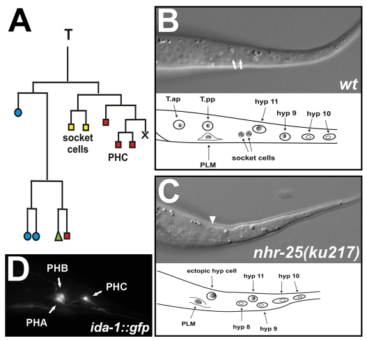
T-seam-cell polarity defect in NHR-25-deficient worms. (A) Lineage diagram of the T seam cell. Blue circles indicate cells that fuse with the hyp7 syncytium; green triangle represents a seam cell; yellow squares mark phasmid socket cells, and red rectangles other neurons. X represents programmed cell death. (B,C) Nomarski images of L2 hermaphrodites demonstrating phasmid-socket absent (Psa) phenotype analysis. Socket cells (white arrows) are the most posterior neurons located between the hyp8 (hyp11) cells and the PLM neuron, which has a typical eye-like appearance (B). An ectopic hypodermal cell (arrowhead) appears instead of the socket cells in the nhr-25(ku217) mutant (C). Anterior is to the left, dorsal up. (D) Phasmids A, B and C marked with the ida-1::gfp transgene in wild type. Anterior is to the left.

