Fig. 1.
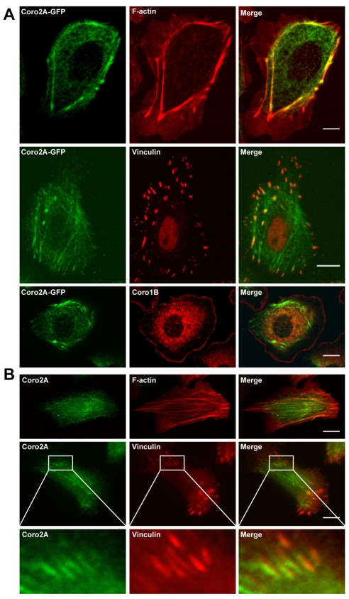
Coronin 2A localizes to F-actin stress fibers and some focal adhesions, but is excluded from the leading edge. (A) MTLn3 cells expressing Coro2A-EGFP were stained with Alexa 568 phalloidin (top panel) or with antibodies against either vinculin (middle panel) or coronin 1B (bottom panel). (B) Immunofluorescence of endogenous coronin 2A (green) with either Alexa 568 phalloidin (top panel) or a vinculin antibody (middle panel). In the bottom panel, magnified images (6×) of the insets in the middle panels show that coronin 2A localizes to focal adhesions. Scale bars: 5 μm.

