Fig. 3.
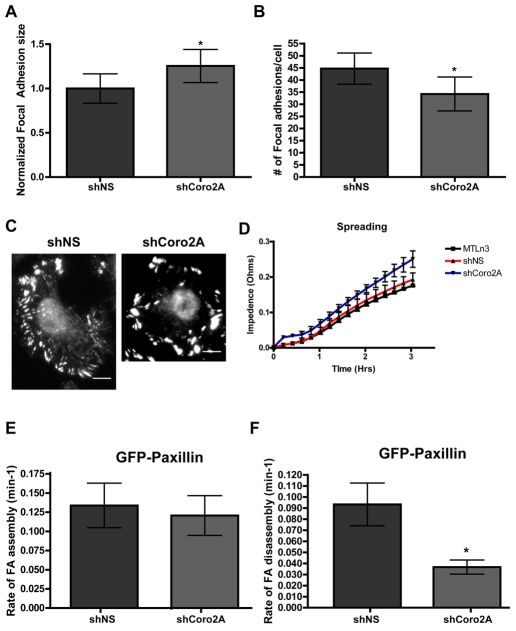
Depletion of coronin 2A increases focal-adhesion size, decreases focal-adhesion number and decreases focal-adhesion disassembly. Focal-adhesion size (A) and number (B) were measured in MTLn3 cells expressing shNS or shCoro2A. Vinculin-positive focal adhesions were used for the quantifications. Focal-adhesion size was normalized against neighboring uninfected cells on the same coverslip. *P=0.0424 for focal-adhesion size and *P=0.0286 for focal-adhesion number, by Student's t-test. (C) Representative vinculin immunofluorescence for MTLn3 cells expressing shNS or shCoro2A used for quantifications in A and B. Scale bar: 5 μm. (D) Cell spreading as measured by change of impedance with the ACEA RT-CES system. Equal numbers of MTLn3, shNS and shCoro2A cells were plated in triplicate. P=0.1726 for MTLn3 versus shCoro2A and P=0.0661 for shNS versus shCoro2A, by Student's t-tests. (E,F) Average rates of focal-adhesion assembly (E) or disassembly (F) visualized by GFP-PXN in cells expressing either shNS or shCoro2A. Cells were imaged once a minute for 30 minutes (see supplementary material Movie 2). Changes of fluorescent intensity were used to determine focal-adhesion assembly and disassembly rates. *P<0.001 by Student's t-test. Error bars represent 95% confidence intervals.

