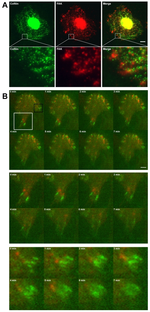
Fig. 7.
Cofilin localizes to the proximal end of some focal adhesions in fixed and live cells. (A) Immunofluorescent images of saponin-permeabilized MTLn3 cells stained for cofilin and FAK. Lower panel shows magnifications (7.5×) of insets. Scale bar: 5 μm. (B) Montage of live-cell images of cell expressing shNS-GFP-PXN and cofilin-TagRFP. Images were taken every minute (see supplementary material Movie 3). Scale bar: 5 μm. Cofilin-TagRFP localizes to the base of GFP-PXN-positive focal adhesions, leading to focal-adhesion disassembly. Middle panel gives enlarged (2.2×) view of white box inset. Bottom panel gives enlarged (4.3×) view of black box inset.

