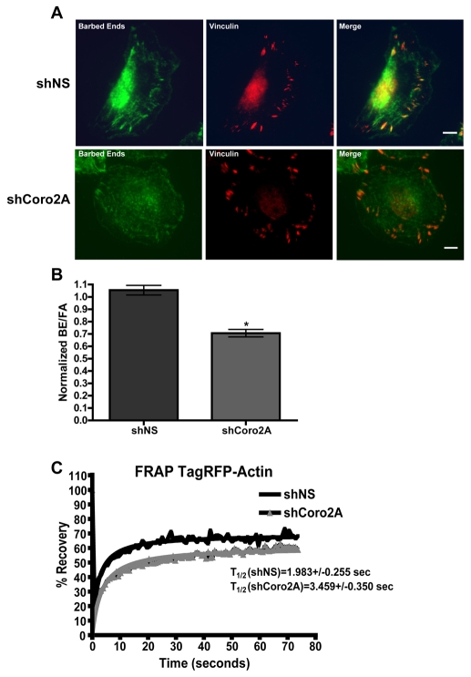
Fig. 8.
Depletion of coronin 2A leads to reduced barbed-end density and actin turnover at internal focal adhesions. (A) Representative images of Oregon green actin conjugate incorporated into free barbed ends at vinculin-stained focal adhesions in MTLn3 cells expressing shNS-TagRFP-actin or shCoro2A-TagRFP-actin. Scale bars: 5 μm. (B) Graph depicting the ratio of barbed ends to focal adhesions (BE/FA), as measured by average fluorescence intensities. These results were normalized to the intensity values of uninfected MTLn3 cells on the same coverslip. Error bars indicate 95% confidence intervals. *P<0.001 by Student's t-test. (C) Graph of fluorescence recovery after photobleaching (FRAP) of TagRFP-actin at focal adhesions in MTLn3 cells expressing either shNS or shCoro2A. T1/2 values indicate time required for 50% recovery of fluorescence. Values from nine cells were used to calculate the recovery rate.

