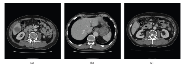
Figure 1.
Abdominal computed tomography. Computed tomography revealed longest diameter 40 mm-sized hepatic mass in the posterior inferior segment of the liver (a) and small nodules scattered throughout both hepatic lobes (b). After 6 courses administration of amrubicin as a third-line chemotherapy, the mass decreased (c).

