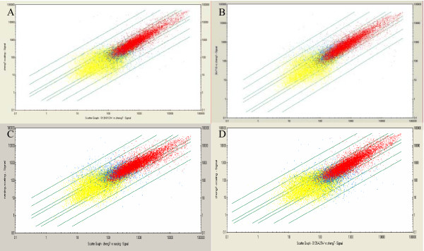
Figure 4.
Scatter plot of gene expression comparisons between the normal rats and DEN-exposured rats. Each point represents a single gene or EST. x-axis: control (from liver tissue of normal rat); y-axis: liver tissue from DEN- treated rat at 12th week (A); at 14th week (B); at 16th week (C); at 20th week (D). The red points represent 'present' states both in control and DEN exposed; blue points represent 'no present' in either of control and DEN-exposed; yellow points represent 'absent' states both in control and DEN-exposed.

